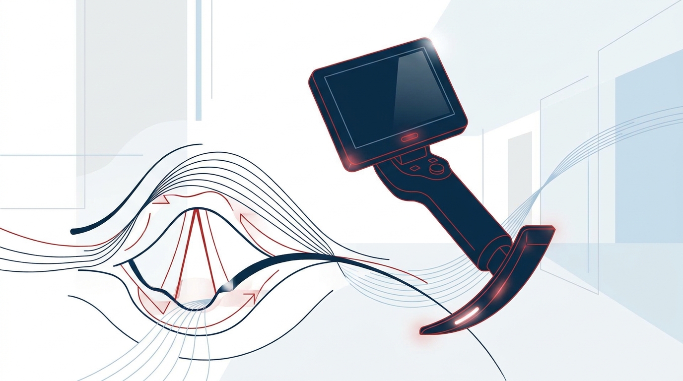
Why video laryngoscopy in emergencies?
Emergency intubation differs fundamentally from elective induction of anaesthesia. Patients are typically not fasted and have limited physiological reserve. There may be blood or vomit in the airway, the cervical spine may be immobilised, and positioning is often unfavourable, for example on the floor, in a vehicle or in poor light. The video laryngoscope shifts the view from the direct line of sight to a screen. The glottis can therefore be seen without having to align the oral, pharyngeal and laryngeal axes.
This gives a practical advantage for the prehospital difficult airway: less force and less head extension are needed to visualise the glottis. The whole team can also see what is happening in the airway. Evidence on hard endpoints such as survival or neurological outcome is limited and depends strongly on the device, the setting and the operator's experience. Video laryngoscopy is therefore a tool, not a substitute for airway competence.
Indications in the prehospital setting and the resuscitation room
Typical situations in which the video laryngoscope offers an advantage
- Anticipated difficult airway: limited mouth opening, short or immobile neck, severe obesity, anatomical abnormalities
- Trauma with cervical spine immobilisation: the glottis can be visualised without relevant head extension.
- Unfavourable patient position: patient on the floor, entrapped or with restricted access to the head
- Resuscitation room and teamwork: the screen supports shared situational awareness, targeted assistance such as external laryngeal manipulation, and structured supervision.
- Cardiac arrest: intubation without prolonged interruption of chest compressions, provided experienced operators are available
Newborns
For preparation, the ERC Guidelines 2025 on newborn resuscitation specify an intubation kit with a video laryngoscope, with the direct laryngoscope as a backup. If the chest does not rise despite optimised mask ventilation, the guidelines recommend suction under direct vision and an increase in pressure. This is followed by a laryngeal mask or intubation with a video laryngoscope, after which the inflation breaths are repeated. Changes to the airway section since 2021 explicitly include the two-person technique, early use of a laryngeal mask and the video laryngoscope [142].
After ROSC
According to ERC/ESICM 2025, the airway should be secured in comatose patients after return of spontaneous circulation. Intubation should be performed by experienced operators, and capnography is mandatory [3]. The video laryngoscope can facilitate this intubation. It does not replace confirmation of correct tube position.
Blade types: Macintosh geometry vs hyperangulated
The choice of blade determines the entire intubation technique. There are two basic shapes, plus channelled systems.
| Feature | Macintosh-type blade | Hyperangulated blade |
|---|---|---|
| Geometry | as a conventional Macintosh blade | markedly curved |
| Laryngoscopy | direct and indirect possible | indirect only (screen) |
| Tube guidance | often without a stylet; bougie works well | preformed stylet usually required |
| Strength | familiar technique; direct view as fallback | view "around the corner", e.g. with limited head extension |
| Typical weakness | limited with a very anterior larynx | good view, but tube placement may still be difficult |
Channelled blades
With channelled blades, the tube sits in a guide channel. It does not have to be steered separately, but the blade must be aligned precisely with the glottis. Small deviations can direct the tube onto the arytenoid cartilages or into the oesophagus.
Practical recommendation
For intubation with a video laryngoscope in emergency medical services, many services have adopted a Macintosh-type blade as standard. It preserves the familiar technique and allows a switch to direct view if the camera is soiled. Hyperangulated blades are a valuable option in specific situations but require dedicated training. The key point is that the team is confident with the system it carries.
Technique: step by step
- Preparation: preoxygenation, suction to hand, bougie or stylet prepared, capnography connected, Plan B and C (for example a supraglottic airway or front-of-neck airway) stated aloud to the team
- Insertion under direct vision: insert the blade in the midline or, depending on the system, from the right. Watch the oral cavity and tongue directly first, and only then switch to the screen.
- Visualise the glottis, do not maximise it: aim for a complete view of the glottis that does not fill the whole screen. If the glottis sits towards the upper part of the screen, there is room for the tube to pass.
- Insert the tube: introduce the tube into the mouth under direct vision until its tip appears on the screen. Only then look at the monitor.
- Passing the glottis: with hyperangulated blades, withdraw the stylet slightly once the tube tip has passed the vocal cords. If the tube catches on the anterior tracheal wall, rotating the tube or using a bougie can help.
- Confirmation of tube position: capnography with a persistent waveform, auscultation and clinical signs. The video view alone does not prove correct tube position.
Common pitfalls
"Good view, but the tube won't go in"
The most common error in reasoning is to equate an excellent view of the glottis with an easy intubation. With hyperangulated blades in particular, the camera axis differs markedly from the path the tube has to follow. The result is a perfect view while the tube catches on the arytenoids, the epiglottis or the anterior tracheal wall.
Countermeasures:
- Withdraw the blade slightly to obtain a less optimal but more accessible view
- Shape the stylet to match the blade curvature
- Use a bougie early
Blade inserted too deep
If the blade is advanced too far, the larynx moves to the upper edge of the screen or out of view. The epiglottis may be lifted up and obstruct the laryngeal inlet. Withdrawing the blade slightly often improves the situation.
Injury without direct vision
Operators who look only at the screen too early are passing the tube blindly through the oropharynx. Soft-tissue injuries to the palatal arches, tonsils or pharynx have been described. The tube should therefore be watched directly until it is visible on the monitor.
Blood, vomit, fogged optics
Contamination is the Achilles' heel of the camera in emergencies. Effective measures include powerful suction, a structured suction technique such as SALAD where appropriate, anti-fog measures and pre-warming of the optics, depending on the manufacturer. In these situations a Macintosh-type blade has the advantage of allowing a switch to direct view.
Prehospital conditions
- Sunlight and glare make the screen hard to read. Shading the screen helps.
- Cold promotes fogging and shortens battery life.
- Equipment checks should be routine: battery, spare blades, function test.
Fixation and loss of time
A video laryngoscope can tempt operators to prolong attempts because "something is visible". Oxygen saturation, not the image, sets the time limit. Criteria for abandoning an attempt and moving to Plan B should be agreed within the team beforehand.
Training and competence
Video laryngoscopy complements conventional laryngoscopy but does not replace it. For emergency physicians and anaesthesia teams this means:
- regular training on the device actually carried, ideally including realistic simulation
- maintaining competence in direct laryngoscopy and supraglottic airway devices
- clear airway algorithms that are communicated within the team
The choice of system, blade shape and local algorithms should follow current airway guidelines and reflect the realities of the respective emergency medical service or resuscitation room.
Practical summary
Video laryngoscopy in emergencies offers clear practical advantages in the anticipated or unanticipated difficult airway, with cervical spine immobilisation and for team communication. It is firmly established in the newborn algorithm of the ERC Guidelines 2025. However, a good view is only half the intubation. Blade geometry, tube guidance, suction and consistent use of capnography determine success.
References
- [142] Hogeveen M, Monnelly V, Binkhorst M et al. European Resuscitation Council Guidelines 2025 Newborn Resuscitation and Support of Transition of Infants at Birth. Resuscitation 2025;215 Suppl 1:110766.
- [3] Nolan JP, Sandroni C et al. European Resuscitation Council and European Society of Intensive Care Medicine Guidelines 2025 Post-Resuscitation Care. Resuscitation 2025 / Intensive Care Med 2025;51:2213–2288.
- [1] Soar J, Böttiger BW et al. European Resuscitation Council Guidelines 2025 Adult Advanced Life Support. Resuscitation 2025;215 Suppl 1:110769 (Korrigendum 2026).
- [4] Kurzfassung der Leitlinien des European Resuscitation Council 2025. Notfall Rettungsmed 2025.
This article does not replace clinical judgement in the individual case or the current summary of product characteristics and guidelines.