Why the stability question comes first
The tachycardia algorithm of the ERC Guidelines 2025 does not begin with the rhythm diagnosis. It begins with the clinical assessment. In the prehospital setting it is often impossible to determine reliably whether a broad-complex tachycardia is ventricular or supraventricular in origin. Whether the arrhythmia is putting the patient's life at risk, however, can be assessed within seconds.
Basic measures following the ABCDE approach:
- Monitoring with ECG, blood pressure and pulse oximetry; apply defibrillator pads early
- Record a 12-lead ECG whenever the situation allows, including before treatment
- Establish IV access and give oxygen as required (target values according to the current guideline)
- Search for reversible causes and treat them in parallel, such as electrolyte disturbances, hypoxia, hypovolaemia, fever, pain or medications
Unstable: criteria and consequences
According to the ERC, the following are considered adverse features:
- Shock: hypotension, peripheral vasoconstriction, altered level of consciousness
- Syncope
- Myocardial ischaemia: chest pain, signs of ischaemia on the ECG
- Heart failure: e.g. pulmonary oedema
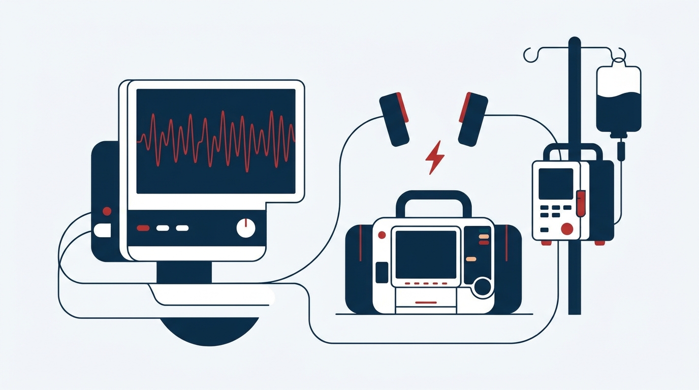
If at least one of these features is present and the tachycardia is the likely cause, synchronised cardioversion is the treatment of choice. This applies regardless of QRS width. A compensatory sinus tachycardia, for example in haemorrhage, sepsis or hypoxia, must be distinguished from this. It is not cardioverted but treated by addressing the underlying cause.
If cardioversion is unsuccessful, the ERC Guidelines 2025 recommend antiarrhythmic therapy (usually amiodarone) followed by repeat cardioversion. Please refer to the current guideline for dosing and the number of attempts.
Synchronised cardioversion in practice
Preparation
- Procedural sedation and analgesia or general anaesthesia by experienced staff. In unstable patients, titrate induction agents cautiously and anticipate cardiovascular collapse. Airway management equipment and a vasopressor should be ready.
- Apply the pads in the anterolateral or anteroposterior position.
- Select a lead with a clearly identifiable, sufficiently tall R wave.
Procedure
- Activate synchronisation mode and check that every R wave is marked. Many devices revert to asynchronous mode after each shock. Therefore re-synchronise before every further shock.
- Select the energy according to the ERC algorithm and the manufacturer's instructions. Please refer to the current guideline or the device manual for specific values.
- Keep the shock button pressed until the shock is delivered on the next R wave. The delay is inherent to the system.
Special situations
- Polymorphic VT or torsade de pointes: reliable synchronisation is often not possible. If the patient is unstable, defibrillate, i.e. deliver an unsynchronised shock.
- Degeneration into ventricular fibrillation: deactivate synchronisation, defibrillate and resuscitate according to the ALS algorithm.
Stable: rhythm analysis by QRS width and regularity
In the absence of adverse features, there is time for a 12-lead ECG and a structured analysis. The tachycardia algorithm differentiates according to two criteria:
- Narrow or broad QRS complex (threshold according to the ERC algorithm)
- Regular or irregular rhythm
Throughout this phase, continuous monitoring is essential. A stable tachycardia can become unstable at any time.
Narrow-complex tachycardia – regular
This is frequently an AV nodal re-entry or AV re-entry tachycardia, less commonly atrial flutter with fixed conduction. The ERC 2025 recommends the following sequence:
- Vagal manoeuvres, preferably using the modified Valsalva technique.
- Adenosine as a rapid bolus followed by a flush, with continuous ECG recording. Even if the tachycardia does not terminate, the transient AV block may unmask atrial flutter.
- If unsuccessful or in case of recurrence: non-dihydropyridine calcium channel blocker or beta-blocker, in accordance with the guideline and contraindications.
Please refer to the current ERC guideline or the summary of product characteristics for dosing. Prepare patients for the brief, unpleasant effects of adenosine.
Narrow-complex tachycardia – irregular
This is usually atrial fibrillation. In stable patients, rate control takes priority, according to the ERC usually with a beta-blocker. In heart failure, alternatives such as amiodarone or digoxin may be considered in line with the guideline.
Rhythm control requires an assessment of duration and thromboembolic risk. This decision should generally be made by cardiology.
Broad-complex tachycardia – regular
A regular broad-complex tachycardia is treated as ventricular tachycardia (VT) until proven otherwise. This applies particularly in patients with known structural heart disease or after myocardial infarction.
- Antiarrhythmic according to ERC 2025: usually amiodarone, dosing according to the guideline or the summary of product characteristics.
- Elective synchronised cardioversion under procedural sedation and analgesia: an option even in stable patients, provided the expertise and setting are available.
- Confirmed SVT with bundle branch block: manage as for narrow-complex tachycardia; adenosine is then an option. In case of diagnostic uncertainty, however, the VT approach applies.
Lidocaine as an alternative
For life-threatening VT with a pulse, the summary of product characteristics specifies the following regimen:
| Step | Dosing |
|---|---|
| Bolus | 1–1.5 mg/kg IV slowly, no more than 25 mg/min (100 mg over ≥ 4 min, 150 mg over ≥ 6 min) |
| If ineffective | Repeat 1–2× after 5–10 min |
| Subsequent continuous infusion | 1 mg/min, up to max. 4 mg/min; in total no more than 200–300 mg per hour |
| Worked example, 70 kg | 70–105 mg = 7–10.5 ml of the 1 % solution (10 mg/ml) |
Source: summary of product characteristics, lidocaine hydrochloride.
Practical notes on lidocaine:
- Check the solution: use only solutions without added adrenaline. Both 1 % and 2 % solutions are available; check the ampoule before administration.
- Dose reduction: in shock, overt heart failure, hepatic impairment or severe renal impairment, approx. 50 % of the dose. Older patients require lower doses.
- Contraindications (outside cardiac arrest): including second- or third-degree AV block, sick sinus syndrome or severe bradycardia, allergy to amide local anaesthetics. In cardiogenic shock, weigh benefit against risk.
- Signs of overdose: PR prolongation or QRS widening.
- Local anaesthetic systemic toxicity (LAST): perioral paraesthesia, metallic taste, agitation, seizures, arrhythmia up to asystole. Treatment includes, among other measures, 20 % lipid emulsion.
- Accumulation during continuous infusion: monitor clinical status and ECG closely.
- Principle: in unstable VT, synchronised cardioversion takes priority, not the drug.
Broad-complex tachycardia – irregular
Three differential diagnoses should be considered.
Atrial fibrillation with bundle branch block
- Manage as for irregular narrow-complex tachycardia.
Pre-excited atrial fibrillation (WPW)
- Recognisable by markedly varying QRS morphology and very high rates.
- Avoid AV nodal blocking agents: adenosine, verapamil, diltiazem, digoxin, beta-blockers. They may facilitate conduction via the accessory pathway.
- If unstable, cardiovert; otherwise consult cardiology.
Polymorphic VT or torsade de pointes
- Consider underlying QT prolongation.
- According to ERC 2025: magnesium sulfate 2 g IV over 10 min, repeat once if required. 2 g corresponds to approximately 8 mmol Mg²⁺.
- Check the concentration on the label. Dilute the 50 % solution before IV administration.
- Magnesium is effective even without measurable QT shortening.
- Discontinue QT-prolonging drugs and correct potassium to the high-normal range.
- In unstable torsade, defibrillate. If pulseless, resuscitate according to the ALS algorithm.
Key points for clinical practice
- Clinical assessment first, then ECG: shock, syncope, myocardial ischaemia or heart failure lead to synchronised cardioversion under procedural sedation and analgesia.
- Check synchronisation before every shock: in torsade or ventricular fibrillation, defibrillate unsynchronised.
- If in doubt, treat broad-complex tachycardia as VT.
- Pre-excited atrial fibrillation: no AV nodal blocking agents.
- Doses of adenosine, amiodarone, beta-blockers and calcium channel blockers as well as cardioversion energies must always be verified against the current ERC guideline and summary of product characteristics.
- Evidence: for many individual steps of the algorithm it is limited and partly based on expert consensus. Local protocols and early involvement of cardiology remain important.
References
- Soar J, Böttiger BW et al. European Resuscitation Council Guidelines 2025 Adult Advanced Life Support. Resuscitation 2025;215 Suppl 1:110769 (Korrigendum 2026).
- Kurzfassung der Leitlinien des European Resuscitation Council 2025. Notfall Rettungsmed 2025.
- Fachinformation Lidocainhydrochlorid 1 % / 2 % Injektionslösung (Antiarrhythmikum) – aktuelle Fassung (fachinfo.de).
This article does not replace clinical judgement in the individual case or the current summary of product characteristics and guidelines.