Why capnography is indispensable in emergencies
Capnography measures the partial pressure of CO₂ in respiratory gas and displays it as a waveform over time. The end-tidal value (etCO₂) depends on three variables:
- CO₂ production (metabolism, temperature)
- CO₂ transport to the lungs (cardiac output, pulmonary blood flow)
- CO₂ elimination (ventilation, airway patency, dead space)
If the etCO₂ value changes, at least one of these variables has changed. This defines the practical value of capnography in emergencies: a single sensor monitors airway, ventilation and circulation continuously and without delay. According to the ERC Guidelines 2025, waveform capnography is an integral part of advanced airway management during resuscitation and of post-resuscitation care. In the post-resuscitation phase, it is explicitly described as mandatory once the airway is secured.
What matters is the waveform, not the number alone. An isolated numerical value without a plausible waveform cannot be relied upon.
Interpreting the capnography waveform: the normal trace
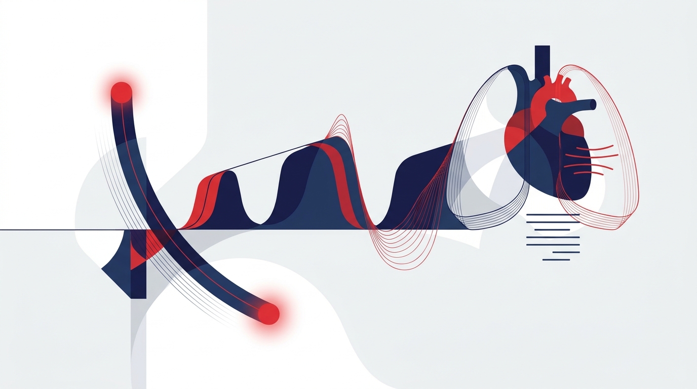
When interpreting a capnography waveform, assess the shape first and only then the value. A normal waveform consists of four segments:
- Phase I – baseline: exhalation of CO₂-free dead-space gas. The line should sit at zero.
- Phase II – steep upstroke: mixture of dead-space and alveolar gas.
- Phase III – alveolar plateau: almost horizontal or slightly rising. Its end point corresponds to the etCO₂.
- Phase 0 – steep downstroke: start of inspiration with CO₂-free fresh gas.
Typical waveform patterns and their significance
- Sudden flat line: disconnection, tube displacement, complete obstruction, equipment failure, or in the worst case cardiac arrest without chest compressions.
- Gradual fall in etCO₂: hyperventilation, falling cardiac output, hypothermia, increasing dead space (e.g. pulmonary embolism).
- Gradual rise in etCO₂: hypoventilation, increasing CO₂ production (e.g. fever).
- "Shark fin" (slurred upstroke, absent plateau): obstructive ventilatory disorder, e.g. bronchospasm, or a kinked or blocked tube.
- Notch in the plateau ("curare cleft"): the patient breathing spontaneously against the ventilator, for example as neuromuscular blockade or sedation wears off.
- Elevated baseline: CO₂ rebreathing, e.g. due to a faulty valve, exhausted absorber or excessive apparatus dead space.
- Cardiogenic oscillations: small ripples in the plateau at low respiratory rates. They are usually harmless.
Confirming tube position with capnography
According to ERC 2025, waveform capnography is the most reliable method for confirming and continuously monitoring endotracheal tube placement. Auscultation, chest movement and tube misting are useful adjuncts but are not sufficient on their own.
Practical approach:
- Connect capnography before intubation so that the waveform is visible from the first breath.
- Look for a reproducible, consistent waveform over several consecutive breaths.
- If the waveform falls away rapidly after initial CO₂ detection, this suggests oesophageal placement. After preceding bag-mask ventilation, CO₂ may have entered the stomach and briefly mimic a waveform.
- Practical rule: "No trace = wrong place". If there is no waveform, the tube is misplaced until proven otherwise.
Limitations:
- With very low pulmonary blood flow, for example during resuscitation with inadequate compression quality, after a long no-flow time or in massive pulmonary embolism, the waveform may be very flat despite correct placement. In this situation, direct visualisation with a videolaryngoscope and, where appropriate, ultrasound are helpful.
- Capnography does not distinguish between tracheal and endobronchial placement. Endobronchial intubation therefore still requires clinical assessment, auscultation and checking of insertion depth.
- Capnography remains the key tool for detecting secondary displacement, particularly during repositioning, transport and patient handover.
etCO₂ during resuscitation
The ERC Guidelines 2025 describe several applications of etCO₂ monitoring during resuscitation:
1. Airway control
Capnography confirms the position of an endotracheal tube or supraglottic airway and monitors it continuously. With supraglottic airway devices, leaks can alter the waveform without the device being misplaced.
2. Ventilation rate
The waveform makes every ventilation visible. Hyperventilation is common under stress, and capnography reliably reveals it. The recommended ventilation rate with a secured airway should be taken from the current ERC guideline.
3. Quality of chest compressions
During resuscitation, etCO₂ mainly reflects the pulmonary blood flow generated by compressions. A fall may indicate declining compression quality, for example due to fatigue of the person performing compressions. Changing the person performing compressions is then appropriate. Evidence for specific etCO₂ targets to guide compressions is limited. The trend is more informative than any single value.
4. Indicator of ROSC
A sudden, marked and sustained rise in etCO₂ during ongoing compressions may indicate return of spontaneous circulation (ROSC). According to ERC 2025, this finding does not replace rhythm and pulse checks but supports the decision on whether to pause compressions. In the differential diagnosis, bicarbonate administration can cause a transient rise in CO₂, as can changes in ventilation.
5. Prognosis: caution required
Low etCO₂ values are associated with a poorer prognosis. However, according to ERC 2025, an etCO₂ value should not be used as the sole criterion for terminating resuscitation. Measurement site, airway, ventilation, compression quality, drugs and the underlying cause influence the value considerably. The decision to terminate resuscitation remains an overall assessment.
Capnography after ROSC
After ROSC, the goal shifts from process monitoring to normocapnia. According to the ERC/ESICM Guidelines 2025 on post-resuscitation care:
- The target is a PaCO₂ of approx. 35–45 mmHg.
- etCO₂ often underestimates PaCO₂. The gradient is increased with enlarged dead space, low cardiac output or lung pathology. Ventilation should therefore be calibrated early by arterial blood gas analysis (ABG).
- During hypothermia, there is a risk of hypocapnia because CO₂ production falls.
- Ventilation is lung-protective with 6–8 ml/kg predicted body weight.
- In addition, an SpO₂ of 94–98 % is targeted; hypoxia and hyperoxia should be avoided.
An unexpected fall in etCO₂ after ROSC may indicate re-arrest, a drop in blood pressure or tube displacement. This applies particularly during transport, when pulse and blood pressure are difficult to assess.
Limitations of capnography in the prehospital setting
Capnography is robust but not infallible. Typical pitfalls in the prehospital emergency service include:
- Technical: moisture, secretions or blood in the sensor or sampling line, kinked sampling lines with sidestream devices, cold, vibration and disconnections during repositioning.
- Physiological: etCO₂ is not a substitute for PaCO₂. In shock, pulmonary embolism, COPD or marked dead space, the difference can be considerable.
- Non-intubated patients: measurements via nasal cannula or mask are well suited for trend and apnoea monitoring, for example during sedation or analgesia. However, absolute values are distorted by mouth breathing, leaks and oxygen flow.
- Interpretation errors: a number without a waveform, a waveform without clinical context, or fixation on a single value lead to wrong decisions.
- Evidence base: for many thresholds, for example for guiding compressions or for prognosis, the data are limited. The ERC Guidelines 2025 are therefore deliberately cautious in their wording.
Practical summary
- Connect capnography before every airway intervention and keep it running continuously until handover.
- Assess the waveform shape first, then the value, then the trend.
- An absent waveform after intubation means misplacement until proven otherwise.
- During resuscitation, etCO₂ indicates airway, ventilation, compression quality and possible ROSC. The value alone is not suitable as a basis for terminating resuscitation.
- After ROSC, aim for normocapnia and compare etCO₂ with ABG early.
References
- Soar J, Böttiger BW et al. European Resuscitation Council Guidelines 2025 Adult Advanced Life Support. Resuscitation 2025;215 Suppl 1:110769 (Korrigendum 2026).
- Nolan JP, Sandroni C et al. European Resuscitation Council and European Society of Intensive Care Medicine Guidelines 2025 Post-Resuscitation Care. Resuscitation 2025 / Intensive Care Med 2025;51:2213–2288.
- Kurzfassung der Leitlinien des European Resuscitation Council 2025. Notfall Rettungsmed 2025.
This article does not replace clinical judgement in the individual case or the current summary of product characteristics and guidelines.