Why the search for causes is part of the ALS algorithm
The ERC Guidelines 2025 on Advanced Life Support include the search for reversible causes as an integral part of the algorithm, for both shockable and non-shockable rhythms. In pulseless electrical activity (PEA) and asystole in particular, the prognosis usually remains poor unless the underlying disorder is treated.
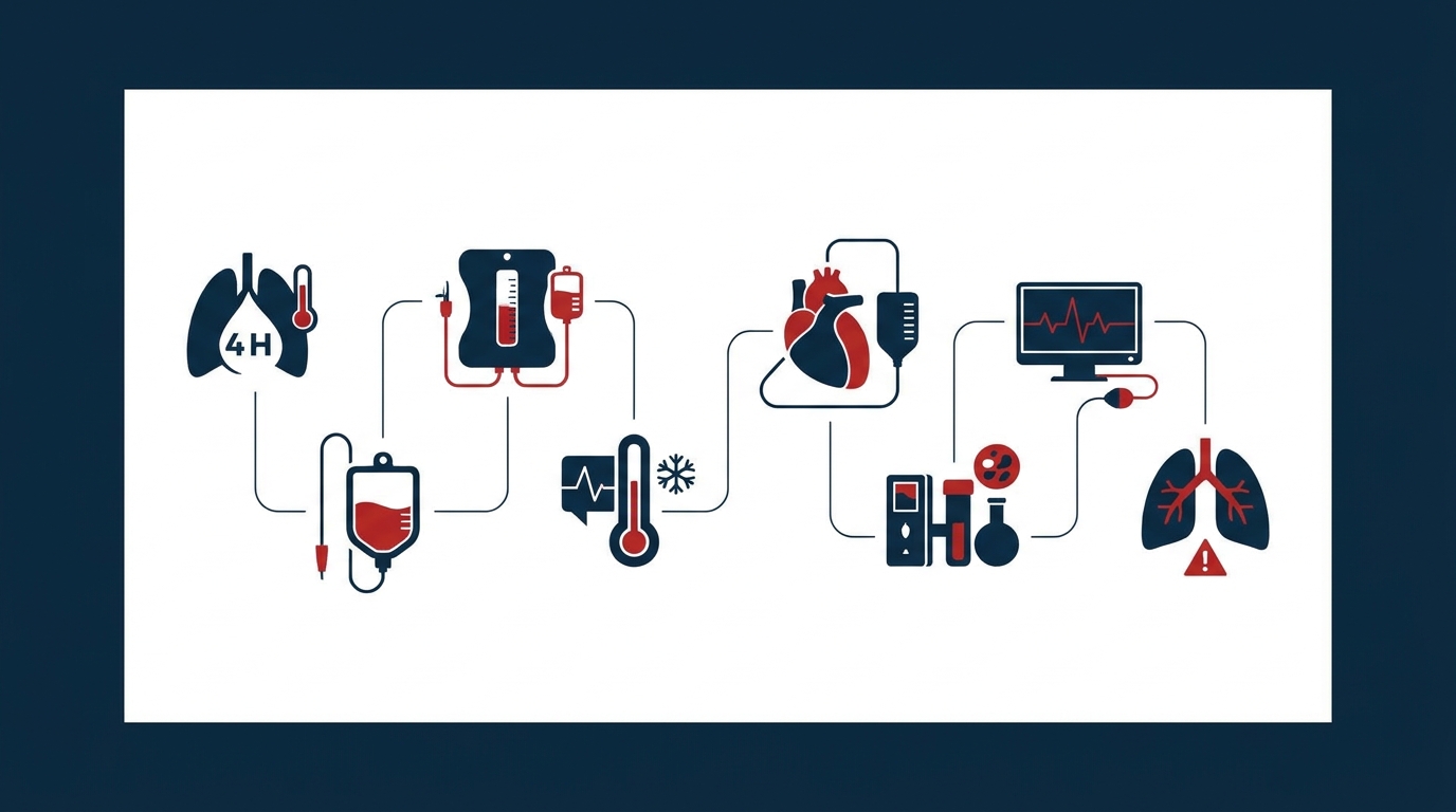
The 4 Hs and 4 Ts mnemonic groups the most common treatable causes of cardiac arrest:
| 4 Hs | 4 Ts |
|---|---|
| Hypoxia | Tamponade (cardiac) |
| Hypovolaemia | Toxins |
| Hypo-/hyperkalaemia and other metabolic disorders | Thrombosis (coronary, pulmonary) |
| Hypo-/hyperthermia | Tension pneumothorax |
The way of working is crucial: the search for causes runs in parallel with resuscitation. It must neither interrupt chest compressions nor delay defibrillation.
Team organisation
It has proven useful to explicitly assign the search for causes to one person, for example the team leader or an additional physician present. The following sources of information should be accessed promptly:
- Context and history: circumstances in which the patient was found, witness accounts, pre-existing conditions, medication, signs of trauma, intoxication or hypothermia
- Clinical examination: chest expansion, neck veins, skin colour, signs of injury, temperature
- Monitoring: capnography, rhythm, etCO₂ trend
- Point-of-care diagnostics: blood gas analysis including potassium, haemoglobin, lactate and glucose; POCUS
A checklist read out loud in every cycle prevents individual causes from being overlooked. It also makes the interim result transparent to the whole team (‘hypoxia treated, potassium pending, tamponade excluded on ultrasound’).
The 4 Hs
Hypoxia
Causes include airway obstruction, drowning, asphyxia and severe respiratory failure. Effective ventilation with a high inspired oxygen concentration is required, together with airway management by experienced providers and capnography to confirm tracheal tube position and to assess the quality of resuscitation. The ERC guidelines also list hypoxia as a treatable cause in the assessment of bradycardia. A hypoxic cardiac arrest may therefore be heralded by progressive bradycardia.
Hypovolaemia
The main causes are haemorrhage (traumatic, gastrointestinal, aortic rupture, gynaecological) and, less commonly, major fluid losses. Treatment consists of haemorrhage control, administration of fluids or blood products, and rapid transport for definitive care. In traumatic cardiac arrest, the ERC Guidelines 2025 recommend addressing reversible causes such as hypovolaemia, tamponade and tension pneumothorax as a priority and simultaneously.
During massive transfusion, citrate from blood products binds calcium. The European trauma guideline recommends monitoring ionised Ca²⁺ and maintaining it within the normal range of approx. 1.1–1.3 mmol/l. Calcium chloride is preferred; the dose is guided by blood gas analysis (Rossaint et al. 2023).
Hypo-/hyperkalaemia and metabolic disorders
Hyperkalaemia is the clinically most important electrolyte disorder in cardiac arrest. Clues include renal failure, dialysis dependence, potassium-sparing medication or preceding ECG changes. Point-of-care blood gas analysis provides a potassium value rapidly.
For cardiac arrest due to hyperkalaemia, the ERC Guidelines 2025 state calcium chloride 10 % 10 ml i.v./i.o. as a rapid bolus (Lott et al. 2025).
- Calcium stabilises the myocardium but does not lower potassium. Additional potassium-lowering measures are needed: insulin–glucose, a β₂-agonist and, if necessary, dialysis. Doses should be taken from the current guideline.
- Calcium chloride 10 % contains about three times as much Ca²⁺ per ml as calcium gluconate 10 %. The ampoule should be checked before administration, as other concentrations are available on the market.
- Do not administer calcium via the same line as sodium bicarbonate without flushing, as precipitation will otherwise occur.
Glucose should be measured in every resuscitation of unclear cause. Other metabolic disorders such as hypokalaemia, hypomagnesaemia or severe acidosis are treated according to the current guideline.
Hypo-/hyperthermia
In accidental hypothermia, rhythm behaviour, drug effects and the likelihood of successful defibrillation change. The ERC guidelines provide modified algorithms for this situation. Transport to a centre offering extracorporeal rewarming (ECLS) should be planned early. Specific temperature thresholds and drug intervals should be taken from the current guideline.
In hyperthermia, for example due to heat stroke or intoxication, rapid active cooling takes priority.
The 4 Ts
Cardiac tamponade
It typically occurs after penetrating chest trauma, after cardiac surgical or interventional procedures, or in aortic dissection. Ultrasound detection of a pericardial effusion with a collapsing right heart can support the diagnosis. Treatment consists of decompression: ultrasound-guided pericardiocentesis or, in traumatic cardiac arrest and with appropriate expertise, resuscitative thoracotomy according to ERC 2025.
Toxins
Clues arise from the circumstances in which the patient was found, the history and the toxidrome. The ERC Guidelines 2025 recommend using specific antidotes where available, contacting a poison information centre early and, in selected cases, considering prolonged resuscitation or ECLS.
- Calcium channel blockers: As an initial dose, the expert consensus states 10–20 ml calcium chloride 10 % or 30–60 ml calcium gluconate 10 % i.v. This is repeated every 10–20 min or given continuously, guided by effect and ionised Ca²⁺, supplemented by high-dose insulin euglycaemia therapy and vasopressors (St-Onge et al. 2017).
- Beta-blockers and calcium channel blockers in the peri-arrest phase: consider glucagon. The dose should be taken from the current toxicology guideline or the summary of product characteristics.
Thrombosis – coronary and pulmonary
Coronary thrombosis: If acute coronary occlusion is suspected, immediate coronary angiography is indicated after ROSC with ST elevation on the 12-lead ECG. In refractory cardiac arrest with a high level of suspicion, mechanical chest compression devices and eCPR may be considered in suitable settings to enable coronary intervention during ongoing resuscitation.
Pulmonary embolism: Clues include risk factors, dyspnoea and syncope before the event, and a PEA arrest. If pulmonary embolism is suspected as the cause, the ERC guidelines recommend considering systemic thrombolysis and then continuing resuscitation for a sufficiently long period. Dose and duration of resuscitation should be taken from the current guideline or summary of product characteristics.
Tension pneumothorax
Typical settings are trauma, positive pressure ventilation, COPD and central venous cannulation. Clinical signs include unilateral absence of breath sounds, distended neck veins and rising ventilation pressures. On ultrasound, absent lung sliding is suggestive. Treatment is immediate decompression, i.e. needle decompression or thoracostomy. In traumatic cardiac arrest, bilateral decompression should be considered according to ERC 2025.
Point-of-care ultrasound in cardiac arrest
POCUS can help identify tamponade, tension pneumothorax, marked hypovolaemia or signs of pulmonary embolism. The ERC Guidelines 2025 attach clear conditions to its use:
- Only by experienced operators: interpretation under resuscitation conditions requires practice.
- No additional pauses in compressions: position the probe before the rhythm check, record a clip during the pause and review it only after compressions have resumed. The pause is determined solely by the rhythm analysis.
- Interpret findings with caution: right ventricular dilatation is common in cardiac arrest and does not by itself prove pulmonary embolism. Absent cardiac wall motion should not be used as the sole criterion for terminating resuscitation.
Transoesophageal echocardiography allows imaging without interrupting compressions. Evidence of a survival benefit is limited so far, and it is available mainly in hospital.
After ROSC: continue the search for causes
Clarifying the cause does not end with ROSC. According to the ERC/ESICM Guidelines 2025 on post-resuscitation care, this includes:
- 12-lead ECG and echocardiography
- laboratory tests including electrolytes, lactate and troponin
- re-assessment of the 4 Hs and 4 Ts
- if the cause remains unclear, whole-body CT (head to pelvis, biphasic, with CT pulmonary angiography)
In parallel, target values are established: SpO₂ 94–98 %, normocapnia and systolic blood pressure above 100 mmHg or MAP above 60–65 mmHg.
Key points for practice
- The 4 Hs and 4 Ts are worked through in parallel in every resuscitation cycle without interrupting compressions.
- Clearly assigned responsibility and a checklist read out loud increase completeness.
- Blood gas analysis and capnography provide important early clues, for example to hyperkalaemia, hypoxia or blood loss.
- POCUS is a tool for experienced operators. It does not replace the overall clinical assessment and must not cause additional pauses.
- In hypothermia, intoxication and pulmonary embolism, prolonged resuscitation and ECLS may be justified.
References
- Soar J, Böttiger BW et al. European Resuscitation Council Guidelines 2025 Adult Advanced Life Support. Resuscitation 2025;215 Suppl 1:110769 (Korrigendum 2026).
- Lott C et al. European Resuscitation Council Guidelines 2025: Special Circumstances in Resuscitation. Resuscitation 2025;215 Suppl 1:110753.
- Nolan JP, Sandroni C et al. European Resuscitation Council and European Society of Intensive Care Medicine Guidelines 2025 Post-Resuscitation Care. Resuscitation 2025 / Intensive Care Med 2025;51:2213–2288.
- Kurzfassung der Leitlinien des European Resuscitation Council 2025. Notfall Rettungsmed 2025.
- Resuscitation Council UK. Special circumstances guidelines – 2025 Resuscitation Guidelines (nationale Adaptation der ERC-Leitlinien 2025), 27.10.2025, Abschnitt Hyperkaliämie.
- Rossaint R, Afshari A, Bouillon B et al. The European guideline on management of major bleeding and coagulopathy following trauma: sixth edition. Crit Care 2023;27:80.
- St-Onge M, Anseeuw K, Cantrell FL et al. Experts consensus recommendations for the management of calcium channel blocker poisoning in adults. Crit Care Med 2017;45(3):e306–e315.
This article does not replace clinical judgement in the individual case or the current summary of product characteristics and guidelines.